
The sine wave ECG is one of the most common types of ECG. It is also called the standard ECG and is used by doctors to diagnose heart disease, arrhythmias and other heart problems.
The sine wave ECG is a type of non-invasive electrocardiogram that is done to measure the electrical activity in your heart. The heart functions as an electrical muscle and sends out electrical impulses to control its rhythm and contractions. When these electrical impulses are measured, they appear as waves on an ECG tracing called a waveform.
When you take an ECG test, you will be asked to lie down on a flat surface while wearing a comfortable shirt or gown so that electrodes can be attached to your body with sticky patches or gel pads. You will also be asked to relax while the technician attaches two small stickers (electrodes) on each side of your chest directly over your heart and another two on both arms just below the elbow joint.
Sine wave ECG
A Sine Wave ECG is a recording of the electrical activity of the heart. The shape of each waveform corresponds with the location of the heart muscle at that moment in time. The heart has four chambers: two atria (upper chambers) and two ventricles (lower chambers). The sine wave refers to the shape of the waves on the ECG tracing. A sine wave is formed when a point on a circle moves in a straight line from one point to another, then returns to its starting point. That is what happens when an electrical impulse travels through an individual heart chamber.
What is sine wave ECG?
Sine wave ECG is a type of ECG that shows a waveform with a simple up-and-down pattern. It is sometimes called a baseline waveform, because it represents the normal electrical activity of the heart in between beats. A sine wave ECG is sometimes used to monitor people who have been diagnosed with an irregular heartbeat or other cardiac conditions.
Sine Wave ECG Recording
A sine wave ECG recording is made by placing electrodes on the chest and shoulders of a patient to record their heart rate and any unusual activity. The electrodes are connected to a machine that records electrical activity from the heart. When you place your finger on your wrist, you can feel your pulse easily because your carotid artery is near your wrist. This artery carries blood from your heart to your brain and head, so if you feel for this pulse, you’re actually feeling blood flow through this artery in your neck.
The same thing happens when doctors take an electrocardiogram (ECG) — they use electrodes attached to the skin of patients’ arms and legs to measure electrical impulses generated by the heart muscle cells. The electrodes are connected to machines that record these impulses onto charts called electrocardiograms or EKGs.
An EKG
Sine wave ECG is a type of ECG that has a smooth, continuous tracing that resembles a sine wave. Sine wave ECGs are usually caused by increased vagal tone on the heart, which slows down the rate of depolarization and results in a slow heart rate.
Sine wave ECGs can be seen in a variety of conditions, including:
Atrial fibrillation (AFib)
Idiopathic ventricular tachycardia (VT)
How do you treat a sine wave?

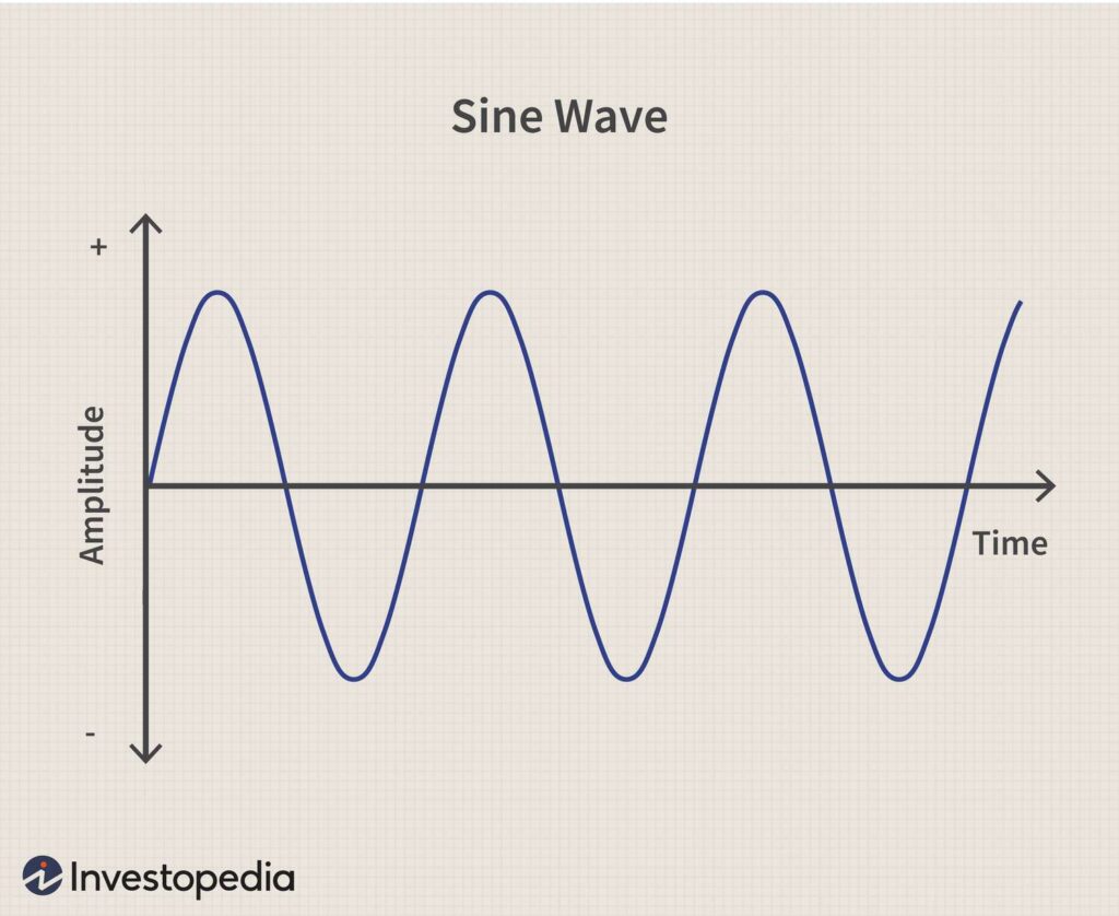
A sine wave is a graph of the function y = sin(x). It is useful in many applications because it can be generated by a pure time-varying voltage source. The voltage of this sinusoid varies smoothly between -1 and +1 volts, but the current through an ideal resistor stays constant at 0 amps.
A sine wave can be represented by its amplitude (a), frequency (f), phase angle (φ), and period T:
y = a sin(2πft)
The amplitude specifies how high or low the wave is above or below its axis. For example, plotting y = 1sin(2πf t) yields a line at an angle of 45 degrees from the x-axis. A positive value for amplitude means that the wave is above its axis; conversely, a negative value means that the wave is below its axis. The phase angle specifies how far ahead or behind from time zero the peak of the sinusoid occurs; again, positive values mean that the peak occurs before time zero; negative values mean that it occurs after time zero. The period specifies how long it takes for one cycle to occur; it can be expressed in terms of seconds (T ) or
Looking at the spectrum of a sine wave, we can see that it has a single frequency component. The spectrum for this sine wave is shown below.
The spectrum for this sine wave has only one frequency component, which is at the fundamental frequency of the sine wave. In general, a pure tone will have a single peak in the spectrum, and all other frequencies in the signal will be zero.
How do you check for hyperkalemia on ECG?
How to check for hyperkalemia on ECG
Hyperkalemia is defined as a serum potassium level greater than 5.5 mmol/l. In the absence of heart disease, it is rare to see levels greater than 6 mmol/l in clinical practice. However, when present, it can cause marked ECG changes that may be difficult to distinguish from other conditions such as acute myocardial infarction (MI).
In an acute situation, where there is severe hyperkalemia, the most common ECG change is peaked T waves. This occurs due to a shift of potassium ions into the extracellular space and away from the intracellular space. As a result, there is less sodium inside the cell and more outside, which causes depolarisation of the cell membrane and therefore repolarisation also occurs more slowly. This results in tall T waves on ECG leads V1-V6 that are usually positive in direction but sometimes negative when associated with ST segment depression.
Other possible ECG changes include widened QRS complexes (QRS widening), shortened PR intervals and prolonged QT intervals (QT prolongation).
The first thing to check is the P wave.
If it’s normal, you don’t have hyperkalemia.
If it’s abnormal, look at the T waves.
If they’re tall and peaked, you have hyperkalemia.
Hyperkalemia can be detected on the ECG by looking for a tall, peaked T wave and prominent U waves. While none of these changes are specific for hyperkalemia, they are consistent with it.
Hypokalemia is indicated by flattened T waves and U waves. Hyperkalemia is indicated by tall, peaked T waves and prominent U waves.
Is sine wave a ventricular tachycardia?
Sine wave is a type of ventricular tachycardia. It is usually a safe rhythm, but can be a sign of complete heart block, or failure of the conduction system.
Sine wave VT is often seen in patients with complete heart block. This type of VT is associated with a slow heart rate and has an abnormal QRS axis (the electrical axis of the heart is abnormal). The QRS complex is flattened out and looks like a sine wave (hence the name). The QRS may have a different configuration from normal, but it does not have to be completely flat.
Sine wave VT may also occur in patients who have had their AV node blocked by an old MI or severe electrolyte abnormalities (hypokalemia or hypomagnesemia).
Sine wave is a type of ventricular tachycardia. It is a regular and abnormal heartbeat that causes an irregular pulse rate. The heart beats at a rate of 100 to 250 beats per minute. This is much faster than the normal heart rate of 60 to 100 beats per minute.
Sine wave is also known as idioventricular rhythm (IVR) or paroxysmal supraventricular tachycardia (PSVT). It occurs in people with heart disease or heart defects.
Sine wave is not a ventricular tachycardia, but it could be an atrial flutter.
Sine wave is usually seen in sinus rhythm with a slow heart rate. Sine waves can also be seen in atrial fibrillation, but the rate will be much faster than in sinus rhythm.
I would recommend that you see your doctor and get a full examination done to rule out any other possible causes of this problem.
Why is it called a sine wave?
The word “sine” is used to describe any mathematical function that produces a smooth curve when plotted on a graph. The word comes from the Latin sine, meaning “without.”
The basic shape of a sine wave looks like the letter S. It goes up and down, with each peak and valley forming an angle with the horizontal axis. The peaks and valleys are evenly spaced, which gives the wave its characteristic shape.
Sine waves are often used in physics because they are simple to calculate and easy to understand, but they also occur in nature — for example, when you throw a stone into a pond or when light reflects off an object. A sine wave also describes how sound waves travel through air or water: They move back and forth at regular intervals.
In mathematics and physics, a sine wave or sinusoid is a mathematical curve that describes a smooth repetitive oscillation. A sine wave is a continuous wave. It is named after the function sine, of which it is the graph. It occurs often in pure and applied mathematics, as well as physics, engineering, signal processing and many other fields. Its most basic form as a function of time (t) is:
y = A sin ( ω t + ϕ ) , {\displaystyle \ y=A\sin(\omega t+\phi ),}
where A represents the amplitude, ω represents the angular frequency (2πf), and f represents the frequency.[1] The wave can be represented as an equation:
y = A sin ( ω t + ϕ ) . {\displaystyle \ y=A\sin(\omega t+\phi ).}
What is an example of a sine wave?
A sine wave is a mathematical function which describes a smooth repetitive oscillation. It can be used to model many naturally occurring oscillations, such as those of a pendulum and ocean waves. The most common form of a sine wave is a pure tone, in which case it’s called a “sine tone”. Sine waves are the building blocks of all other waves, such as those that make up music or sound effects.
Sine Wave Examples
The most common example of a sine wave is a pure tone. If you sing into your bathroom mirror at home, you will hear your voice as a series of pure tones. These pure tones are sine waves:
A pure tone consists of one frequency (pitch) only — there are no overtones or harmonics present. Pure tones are often generated by musical instruments like pianos and violins, but can also be produced by other sources such as electrical circuits with passive components (capacitors) or mechanical devices like drum sets and pipes.
Another example of a sine wave is the sound made by wind chimes when they’re blown by the wind — this is also known as an aerodynamic whistle:
The frequency of the sine wave is the number of cycles it completes in one second. The amplitude is how far above or below the rest position each cycle reaches.
A sine wave can be generated by moving an object back and forth through space at a constant speed, or by compressing and expanding a spring at regular intervals. These are called mechanical waves because they are produced by physical movements — not by electricity or magnetism.
What is considered ventricular tachycardia?
Ventricular tachycardia (VT) is a fast heart rate that starts in the heart’s ventricles. It’s the most common type of rapid heartbeat.
Ventricular tachycardia can be dangerous and may lead to sudden cardiac arrest if not treated quickly. In general, ventricular tachycardia is less common than supraventricular tachycardia in people with heart disease, but it tends to be more serious because it can cause sudden cardiac arrest.
Ventricular tachycardia is caused by a problem with the electrical signals that control your heartbeat. These signals travel down pathways in your heart called Purkinje fibers and bundle of His fibers, which are bundles of specialized muscle cells in your heart’s upper chambers (ventricles). The signals travel through these fibers and cause contraction of your ventricles’ muscle tissue — which pumps blood out of your heart.
If you have an abnormal pattern of electrical activity in your ventricles, you may have ventricular tachycardia.
Ventricular tachycardia (VT) is an abnormally fast heart rhythm that originates from the ventricles. It is a type of supraventricular tachycardia, which means it originates above the ventricles in the atria. It can lead to serious health complications if not treated immediately.
Ventricular tachycardia occurs when electrical impulses originating in the ventricles begin firing faster than normal. This causes the ventricles to contract too quickly, leading to a higher heart rate and blood pressure. The condition is sometimes confused with ventricular fibrillation (VF), which causes erratic electrical activity in the ventricles that can result in sudden death.
What are the types of ventricular tachycardia?

Ventricular tachycardia is a rapid heart rate that begins in the ventricles, or lower chambers of the heart. The most common type of ventricular tachycardia is ventricular fibrillation.
Types of Ventricular Tachycardia
There are three major types of ventricular tachycardia:
Paroxysmal (or paroxysmal supraventricular) tachycardia (PSVT) is the most common type of SVT. It’s short-lived and generally doesn’t cause any symptoms or problems. PSVT can occur at rest or during exercise; it usually goes away within a few minutes without any treatment. If you have PSVT, you may experience palpitations and lightheadedness, but these feelings usually go away quickly on their own without treatment.
Persistent (or permanent) forms require treatment because they can lead to more serious heart rhythm problems like sudden cardiac death. Persistent paroxysmal supraventricular tachycardia (PPVST) is a form of PSVT that lasts longer than 10 seconds and returns after being stopped with medication or electric shock therapy (cardioversion). Persistent atrial fibrillation (AFib) is another form
Ventricular tachycardia (VT) is an abnormal heart rhythm that originates in the ventricles. It’s typically caused by a disturbance in the electrical system of the heart, which causes it to beat very fast.
There are two types of VT — narrow-complex and broad-complex.
Narrow-complex VT: This type of VT involves a normal QRS complex (the waveform on an electrocardiogram that represents depolarization of the ventricles). Because this type of VT has a normal QRS complex, it’s sometimes referred to as narrow-complex tachycardia (NCT). Narrow-complex VT is usually less serious than broad-complex VT, but can still be life threatening if not treated quickly.
What types of waves are sine waves?
sine waves are a special type of wave. They are the simplest type of wave, and they occur naturally in many systems. Sine waves can be found in the ocean, sound waves, and electricity.
The term “sinusoid” was coined by German mathematician Gottfried Leibniz in 1694; it is derived from the Latin sinusoidum, which translates to “like a sine.” The name is appropriate because sine waves closely resemble the shape of an arch or curve formed by the sines of trigonometric functions.
The term “sine wave” is also used loosely to describe other types of periodic waveforms that have no mathematical relationship to sinusoids. For example, sawtooth waves are not true sinusoids because their amplitude does not decrease smoothly with increasing frequency as do true sine waves.
Sine waves are a type of waveform. They’re periodic functions with a smooth continuous curve, which means they have no sharp corners or sudden changes in value.
In math and physics, sine waves are often used as an approximation to more complex waveforms, such as a square wave or sawtooth wave. Sine waves are also often used in signal processing and audio applications.
Sine waves are typically described by the amplitude (or height) of their peaks and troughs, the wavelength (or distance between peaks), and the frequency (or number of cycles per second).
What is the difference between SVT and St?
The major difference between SVT and St is that SVT is a narrow-complex tachycardia (the heart rate is fast) that occurs due to abnormal electrical impulses in the atria, while St is a broad-complex tachycardia (the heart rate is fast) that occurs due to abnormal electrical impulses in the ventricles.
SVT is generally caused by paroxysmal supraventricular tachycardia (PSVT), which means the cause of the abnormal electrical activity is above the ventricles. PSVT can be caused by a re-entrant circuit or an accessory pathway. The latter may be congenital or acquired, such as during pregnancy.
Strep throat, however, is a bacterial infection of the throat that causes inflammation and sometimes scarring of mucous membranes.
SVT is a group of heart arrhythmias that can cause rapid heartbeat and palpitations. The most common kind of SVT is atrial fibrillation, or AFib.
St is an abnormal heart rhythm caused by an electrical disturbance in the upper chambers (atria) of the heart.
SVT is usually easy to treat with medication and sometimes a procedure called cardioversion, which restores normal sinus rhythm.
St requires long-term treatment with medications or an implantable cardioverter defibrillator (ICD).
The main difference between SVT and St is that SVT is a problem with the electrical system of the heart, while ST is a problem with the heart’s electrical system.
SVT stands for supraventricular tachycardia, while St stands for sinus tachycardia.
Both are types of arrhythmias, which are disturbances in the normal rhythm of the heart’s beating. But they are different from each other in their causes and symptoms.