
The uvula deviates to the right in about 90% of people.
The uvula is a small, fleshy, somewhat triangular extension from the posterior border of the soft palate. It hangs down into the throat and may be visible when you open your mouth wide.
The soft palate is made up of two flaps of mucous membrane that stretch from just behind the nose to the back wall of the throat. The uvula is attached to one of these flaps.
When you swallow, your uvula moves upward and backward to protect your airway from food or drink going down into your lungs. If you have ever seen someone with an obstructed airway (for example, someone having difficulty breathing), they may have a swollen or inflamed uvula that looks like a small balloon on their soft palate. This happens because they are having trouble swallowing properly and their saliva is building up in their mouth instead of going down into their stomachs.
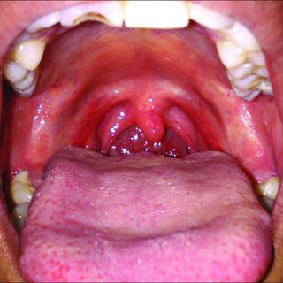
The uvula deviates toward the right side. The uvula is a small inverted V-shaped structure that hangs down from the posterior part of the soft palate. It forms the posterior boundary of the nasopharynx, which is located behind and below the nasal cavity.
The uvula forms from a primary outgrowth from the palate during development from around week 4 of gestation. It grows towards the midline and then loops back posteriorly to reach its final position in front of the soft palate.
The velopharyngeal port is formed by two muscles: levator veli palatini (LVP) and tensor veli palatini (TVP). LVP is responsible for moving palatal tissue posteriorly, which opens up a space for air to travel through during speech production. TVP acts to tense this same tissue, thereby closing off this space and preventing food from entering it during swallowing.
What nerve causes uvula deviation?
The uvula is a small piece of soft tissue that hangs from the back of your throat. When you’re talking or eating, the uvula can move around to block the nasal passage or the back of your throat. This can cause you to have trouble breathing through your nose, which can be uncomfortable and make it hard to speak clearly.
Uvula deviation (also called uvular deviation) occurs when the uvula moves from its normal position in the back of your throat and hangs down into your mouth.
There are many reasons why people experience uvula deviation, including:
Acid reflux. If you have acid reflux, stomach acid can get into your esophagus and irritate it. This can cause stomach pain and heartburn — along with other symptoms like a sore throat, hoarseness and trouble swallowing.
Overweight or obesity. Being overweight or obese puts pressure on your chest cavity, which can make it difficult for air to pass through your nose when you breathe in deeply (known as inspiratory stridor).
Injury or trauma to the neck or head area. Trauma such as whiplash or a bad bump on the head can cause irritation in this area, resulting in uvula deviation
The glossopharyngeal nerve is a cranial nerve that gives rise to the ninth and tenth cranial nerves. It supplies several muscles in the head and neck, including the stylopharyngeus muscle, which causes the uvula to move up into the nasal cavity when swallowing.
Other muscles innervated by this nerve include:
Taste buds of the posterior two-thirds of the tongue
Taste buds of the pharynx (partially)
Taste buds of upper esophagus (partially)
Does the uvula deviate to the side of the lesion?
The uvula can deviate to either side of a lesion. The uvula is the soft, fleshy structure that hangs down from the back of your palate. It’s the little “thing” at the back of your throat that you sometimes see when you stick out your tongue.
The uvula is attached to the soft palate (the soft tissue at the back of your mouth and throat). The soft palate covers the opening between your mouth and nasal cavity and helps control airflow from the nose into the throat. When you eat or drink, food or liquid goes through this opening into your esophagus (the tube carrying food from your mouth to your stomach). If something blocks this passage, it can cause choking.
The uvula sits right on top of one of these critical areas — where air enters the throat. When something blocks this opening, it can cause choking.
If something blocks this passage, it can cause choking.”
The uvula is a small, fleshy structure that hangs down from the back of the throat. It can have a slight bend in it, which can make it deviate to one side or another, but this is not always present.
The uvula is normally straight and should not be deviated to either side when you are awake and alert. If you are having difficulty swallowing liquids, particularly if you feel as though there is something stuck in your throat, then it may be because there is a foreign body lodged in the back of your mouth or throat.
Foreign bodies in the back of your mouth can include food particles, gum and toothpaste residue, bits of dental work or part of a tooth root (which might have come loose after an extraction).
If there is no obvious cause for this feeling then you should see your doctor to ensure there is nothing serious going on.
Which cranial nerve elevates the uvula?
The glossopharyngeal nerve (CN IX) is the only cranial nerve that elevates the uvula. The vagus nerve (CN X) also innervates the muscles of the larynx, but it does not affect the uvula.
The glossopharyngeal nerve has a general sensory component and a special sensory component. The sensory component contains axons that carry taste sensations to the brain. These axons are located in the posterior part of the lingual nerve (a branch of CN V3), which ascends through a cleft between two muscles called genioglossus and styloglossus (located on either side of Adam’s apple).
When you chew food, these muscles contract simultaneously, causing your tongue to move up and down against your palate. The movement causes friction between your tongue and palate, resulting in an increased sensitivity to taste.
The seventh cranial nerve (the facial nerve) controls most of the muscles involved in facial expression and hearing.
The seventh cranial nerve is responsible for elevating the uvula, a small piece of soft tissue that hangs from the back of your soft palate. The uvula helps to prevent food and liquids from entering your nasal passages when you swallow.
The facial nerve also controls the muscles involved with chewing and swallowing food. In addition, this nerve is responsible for controlling the movement of your eyes as well as closing them when you sleep.
Why is my uvula deviated to one side?
The uvula, which is the dangly thing at the back of your mouth, can be deviated to one side.
The most common cause of an uvular deviation is an injury to the uvula, which can happen if you hit it on something or have a migraine. In some cases, people are born with a deviated uvula.
If you have a deviated uvula, it may be difficult to breathe through your nose and you might have trouble speaking clearly. You may also have bad breath caused by food getting stuck under your tongue and rotting behind your teeth.
The treatment for an uvular deviation depends on its cause and severity. If your uvula is simply crooked and not interfering with breathing or swallowing, there’s no need for treatment unless it bothers you personally (for example if it makes you feel embarrassed). In some cases, treating underlying conditions such as allergies or acid reflux may make the problem go away on its own.
What muscles elevate the uvula?

The levator palatini, a muscle in the soft palate, can elevate the uvula.
The levator veli palatini muscle is the only muscle that elevates the uvula. It originates from the foramen cecum of the palatine bone and inserts into the soft palate. When it contracts, it pulls up on the soft palate and uvula.
The tensor veli palatini muscle extends from the posterior surface of the soft palate to a tendinous band that stretches across its posterior border. It runs alongside another muscle called the levator palatini muscle, which is responsible for raising your uvula.
The tensor veli palatini also helps keep food or liquid out of your nasal cavity by contracting when you swallow or yawn to prevent liquids from entering your nose instead of your mouth.
The uvula is a small tissue that hangs down from the back of your soft palate. It’s usually in your throat when you’re not swallowing, but it can move around.
The uvula is controlled by muscles in your mouth, pharynx, and nasal cavity. The superior pharyngeal constrictor muscle pulls the uvula up toward the soft palate. The palatopharyngeal arches lift the uvula as well.
Adenoids are a large gland near the back of your nose that can get infected and swollen if you have allergies or other problems with your immune system. Adenoids can make it hard to breathe through your nose and cause other problems with breathing too.
What does Cranial nerve XI do?
Cranial nerve XI (the spinal accessory nerve) supplies the sternocleidomastoid and trapezius muscles. It also provides sensory innervation to the skin over the lateral aspect of the head, posterior surface of the external ear, and upper portion of the trapezius muscle.
The spinal accessory nerve arises from the brainstem as a continuation of cranial nerve XII (hypoglossal nerve), which exits from above and behind the jugular foramen at C1-C2. The spinal accessory nerve passes superior to the vertebral artery within its canal in the neck, then leaves this canal to enter a groove on the surface of vertebrae C2-C6. It divides into two branches, separated by an intermediate loop that extends over C4-C5. These branches supply motor innervation to sternocleidomastoid and trapezius muscles; they also provide cutaneous sensation over the lateral aspect of the head and posterior surface of external ear
What happens if cranial nerve 9 is damaged?
If a person’s ninth cranial nerve is damaged, they may experience loss of taste on one side of the tongue.
The nerve also carries signals that connect to the muscles of facial expression, so if it’s damaged, a person may lose some of their ability to move certain facial muscles. This can result in an inability to close one eye or raise one eyebrow, for example.
If the ninth nerve is damaged, it can cause problems with chewing and swallowing. The person may have difficulty moving food from one side of the mouth to the other as they chew, which can lead to improper chewing and eating habits. This can cause problems with processing food and lead them to develop nutritional deficiencies as well as dental issues such as cavities or tooth decay.
Cranial nerve 9 is the oculomotor nerve, which is responsible for moving the eye muscles.
If this nerve is damaged, it can cause double vision and impaired eye movements. This could be due to damage in either the brain stem or the eye muscles themselves.
Cranial nerves are a group of nerves that originate from the brainstem or spinal cord and are responsible for sending messages to different parts of your body. There are 12 pairs of cranial nerves: each pair controls a specific part of your body. For example, cranial nerve 9 (the oculomotor nerve) regulates eye movement and keeps your eyelids closed when you blink your eyes.
How do you test cranial nerve 9?

Cranial Nerve 9 (VII) is the facial nerve. It supplies all of the muscles for facial expression, except for the frontalis muscle, which is supplied by CN 5. The nerve also supplies taste from the anterior two-thirds of the tongue, a portion of the external ear canal and the lacrimal gland.
The VIIth nerve is tested by evaluating facial movement and sensation on the face and scalp.
Anatomy: The main trunk of CN VII exits the skull base through an opening in the petrous portion of temporal bone (petrotympanic fissure). In adults, it travels through ponto-mesencephalic canal, but in fetuses it goes through foramen lacerum instead. There are two divisions, temporal and zygomatic, each with its own motor nucleus (temporal = motor nucleus; zygomatic = motor nucleus). The motor root leaves cranial cavity via hypoglossal canal (anteroinferior to lingual nerve) then passes through trigeminal ganglion en route to muscles innervated by it (except frontalis). It then leaves trigeminal ganglion via foramen ovale and travels into orbit where it branches out into superior orbital fissure
The ninth cranial nerve, called the glossopharyngeal nerve, is a special sensory nerve that provides taste sensation from the posterior third of the tongue and general sensation from the pharynx (back of throat).
The glossopharyngeal nerve has three major branches:
Vagus nerve. The vagus nerve is an important part of the parasympathetic nervous system. It stimulates various internal organs, including those of the digestive tract and heart.
Accessory nerve. The accessory nerve is a motor branch that innervates muscles of the head, neck, and thorax. It also has taste buds in its lingual branch that supply taste to the posterior third of the tongue.
Glossopharyngeal nerve (gag reflex). This branch supplies general sensation to parts of your pharynx (back of throat) and taste buds to your epiglottis (the flap at the opening between your larynx and esophagus). It also plays an important role in triggering vomiting when something goes down your throat but doesn’t belong there
What does glossopharyngeal nerve do?
The glossopharyngeal nerve is a cranial nerve that supplies the posterior third of the tongue, soft palate, pharynx, and carotid body.
The glossopharyngeal nerve (cranial nerve IX) arises from the medulla oblongata, where it originates with fibers from the vagus nerve (cranial nerve X). It passes around the superior cervical ganglion and enters the skull through the jugular foramen. Within the skull it gives off branches to the pharynx and carotid body. In addition to supplying taste sensation to the posterior third of tongue, it also carries general sensory information from this region. It also sends parasympathetic postganglionic fibers to paraganglia in the carotid body that modulate blood pressure.
Glossopharyngeal neuralgia is an uncommon form of trigeminal neuralgia where pain occurs in areas supplied by glossopharyngeal nerves.[1] This condition usually occurs in elderly women.[2]
The glossopharyngeal nerve (CN IX) is the ninth cranial nerve. It supplies motor fibers to the stylopharyngeus muscle and sensory fibers to the posterior third of the tongue, tonsils, pharynx, and carotid body.
The glossopharyngeal nerve is a mixed nerve made up of both sensory and motor components. The sensory component provides sensation in the posterior third of the tongue, tonsillar fossa, pharynx, larynx and external auditory meatus (EAM). It also carries preganglionic parasympathetic fibers from the brainstem to synapse with postganglionic parasympathetic neurons in one or more ganglia within or outside of the head and neck region. The motor portion supplies muscles of stylopharyngeus, palatopharyngeus, styloglossus and tensor veli palatini.[1][2]
Sensory function
The sensory function of CN IX can be divided into two groups: visceral and special sensory functions.[3] Visceral sensation includes taste from the posterior 2/3rds of tongue; pain from posterior 1/3rd of tongue;[4]
The glossopharyngeal nerve (CN IX) is the ninth cranial nerve. It is a mixed nerve that carries both sensory and motor information from the posterior third of the tongue, tonsils, pharynx and carotid body to the brainstem.
The CN IX consists of two separate pathways: a central nervous system portion and a peripheral nervous system portion. The central nervous system portion carries taste sensation from the posterior third of the tongue, while the peripheral nervous system portion carries motor fibers that innervate muscles of the pharynx, larynx and soft palate.
In addition to these functions, the glossopharyngeal nerve also carries afferent fibers from several visceral structures including those in the pharynx (tonsils), heart (carotid sinus), esophagus (cardiac muscle) and lungs (bronchial muscle).
The glossopharyngeal nerve is a cranial nerve that innervates the posterior third of the tongue, the pharynx (which includes the soft palate and palatine tonsils) and the epiglottis.
The glossopharyngeal nerve is a mixed nerve. The sensory component carries taste sensations from the posterior third of the tongue, and general sensation from the pharynx including taste from the posterior third of the tongue. The motor component has visceral efferent fibers that control muscles in the pharynx and larynx, including those involved in swallowing.
The glossopharyngeal nerve is derived from cranial nerve VIII (the vagus nerve), which gives rise to its sensory component, as well as its motor component. The sensory component emerges above the vagus nerve and pierces the carotid sheath to reach its target organs.[1] The motor component exits below this point and travels through an opening in this sheath called a tympanic plexus.[2]
The glossopharyngeal nerve has two roots: an anterior root originating from cranial nerve IX (the accessory nerve), and a posterior root originating from cranial