The 12-lead ECG is a snapshot of the electrical activity of your heart over a period of time. It shows the electrical activity from 12 different positions (leads) around the body.
The 12-lead ECG is one of the most important tests that your doctor can perform when you go to see them with a problem related to your heart or lungs. The ECG is also called an EKG or ECG.
The 12 leads are numbered from 1 to 12, starting at the top left corner and moving counter-clockwise around your body (viewing from above). Each lead has a letter associated with it as well, making it easier to remember where each lead is placed on your chest/back:
Lead I: positive electrode on right leg, negative electrode on left leg;
Lead II: positive electrode on left leg, negative electrode on right arm;
Lead III: positive electrode on left arm, negative electrode on right leg;
Lead IV: positive electrode on right arm, negative electrode on left leg;
The 12 lead ECG is a vital tool in the diagnosis of many cardiac conditions.
The placement of leads on the body is important as it affects the quality of the ECG tracing. The 12 lead ECG has a standardised placement which helps to standardise interpretation by physicians.
The 12 lead ECG is used to diagnose a variety of conditions including:
Myocardial infarction (heart attack)
Congestive heart failure
Pericarditis (inflammation of the pericardium)
Arrhythmias (abnormal heart rhythms)
How do you remember the ECG 12 lead placement?
There are two ways to remember the placement of the ECG leads.
The first way is by thinking about the heart’s electrical axis. This is the axis around which your heart’s depolarization occurs. In other words, it is the direction in which your heart is most likely to depolarize first in a given beat (meaning that it has a high probability of being positive).
There are three possible axes:
- Right axis deviation: The electrical axis passes through the right side of your chest. This means that the right ventricle is most likely to depolarize first during each beat (since it has a higher density of muscle tissue than the left ventricle).
- Left axis deviation: The electrical axis passes through the left side of your chest. This means that the left ventricle is most likely to depolarize first during each beat (since it has a higher density of muscle tissue than the right ventricle).
- Normal axis: The electrical axis passes through your chest at an angle between 0 and 45 degrees, meaning that both sides are equally likely to depolarize first during each beat
- There are 12 leads, each with a letter code. The letters are arranged in a circle and the leads are placed on the chest in this order:
- A-VR I-II III-aVR aVL aVF B-VL II-III aVF V1 V2 V3 C-VF III-aVL aVR V4 V5 V6 D-I aVR I bVL II bVF III+IV
How do you remember where to place leads?
A lot of people ask me how I remember where to place leads. It’s a simple process that takes me about five minutes a day.
I use a whiteboard to keep track of my leads. Each time someone emails me with an interest in my product or service, I put their name on the board. For example, let’s say I have three people interested in buying my premium course.
I’ll write down their name on my board, and draw a dot next to it. If I get another email from one of these people later in the week asking for more information about how to purchase the course, I’ll just circle their name on the board again so that I remember who they are. The next time they contact me again, I’ll cross out their name and move it over to the right side of the board where they’re crossed out because they’ve already been contacted twice but haven’t purchased anything yet (this doesn’t mean they won’t buy but simply means that you need to follow up with them more often).
I’m a designer and I’ve never been able to remember where all my leads should go. This is frustrating because it takes up a lot of time searching for the right lead in order to complete a circuit.
I’ve tried using different color wires and connectors but that doesn’t help much, since I have so many wires in each circuit.
It would be great if there was a way to label all the leads with their respective locations. Or even better, if there was some sort of system that could automatically place leads into their correct location without having to move them manually.
Why is it called a 12 lead ECG when there are only 10 leads?
There are actually 12 leads on the EKG machine. The reason it’s called a 12 lead ECG is because there are 12 electrodes that are attached to your chest, neck and arms. There are 10 electrodes that are used to record the electrical activity of the heart. These electrodes monitor electrical activity from different parts of the heart, so they can be used to determine if there is any damage from a heart attack or other heart problems.
The other two electrodes are really just spares in case one needs to be replaced during testing. If you’re curious about what each lead represents, here’s a quick rundown:
Lead I (Positive Left) – This lead records voltage between your left arm and left leg. It represents depolarization in your AV node and bundle branches.
Lead II (Positive Right) – This lead records voltage between your right arm and right leg. It represents depolarization in your atria and ventricles (the top chambers).
Lead III (Negative Left) – This lead records voltage between your left arm and left leg. It represents repolarization in your AV node and bundle branches.
Lead IV (Negative Right) – This lead records voltage between your right arm and right leg. It represents depolar
What does V1 V2 V3 mean in ECG?
The term V stands for the vertical axis on an electrocardiogram (ECG). The three main parts of the ECG are PQRST, where P and T are horizontal axes. The first number indicates which lead the heart is in (Lead I), second number how many millivolts above or below zero it is (the amplitude), and third number is how many milliseconds after the start of the QRS complex it occurred. The reason for this is that there is no way to represent negative voltages on paper so they are instead represented as positive values which are then subtracted from zero (0) to get a value between 0 – 6.
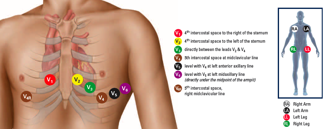
V1: First Intercostal space at right sternal border
V2: Second Intercostal space at left sternal border
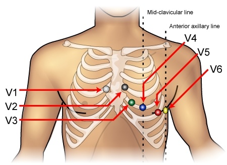
V3: Third intercostal space at left midclavicular line
A standard 12-lead ECG has twelve different leads. Each lead represents a different viewing angle of the heart. The three most common leads are:
V1 – Right arm, positive electrode on the left arm, negative on the right leg
V2 – Left arm, positive electrode on the left leg, negative on the left arm
V3 – Left leg, positive electrode on left leg, negative electrode on right arm
The P wave represents atrial depolarization, and it occurs before every QRS complex. The PR interval is the time from the onset of the P wave to the beginning of the QRS complex. The QRS complex represents ventricular depolarization.
The T wave represents ventricular repolarization. The ST segment is a short segment of the ECG that separates two adjacent T waves. The U wave is a small upward deflection following the T wave in some leads (unipolar leads V2-V6).
How many chest leads are placed for a 12 lead ECG?
12 lead ECGs are made from six combinations of leads, called limb leads, and one combination of chest leads. The limb leads are V1 through V6, and the chest leads are augmented by two more (one lead each) which take advantage of the chest’s electrical properties to provide extra information about the heart’s electrical activity.
The standard 12-lead ECG has eight horizontal leads (I, II, III, aVR, aVL, aVF) and four vertical leads (aVRR’, aVLQ’, aVFR’, AVLQ’). These are named after their corresponding limbs: I corresponds to the right arm (ra), V1 corresponds to the first half of the right arm (rH), and so on.
The other four combinations are:
aVRR’ – augmented right arm
aVLQ’ – augmented left leg
aVFR’ – augmented right foot
AVLQ’ – augmented left foot
The 12 lead ECG contains 12 electrodes:
Lead I: The electrode is placed in the first intercostal space of the left mid-clavicular line.
Lead II: The electrode is placed in the second intercostal space of the right anterior axillary line.
Lead III: The electrode is placed in the third intercostal space of the left anterior axillary line.
Lead aVR: The electrode is placed on the right shoulder, just below the clavicle and at the level of V3 (so it’s really more like V1 or V2).
Lead aVL: The electrode is placed on the left shoulder, just below the clavicle and at the level of V4 (so it’s really more like V5 or V6).
Lead aVF: The electrode is placed on your right hip bone (just above your hip joint), about 2 inches away from your belly button.
What does ECG 12-lead stand for?
ECG stands for electrocardiography. The ECG is a test that records the electrical activity of your heart. Your heart sends out electrical impulses when it contracts, and these impulses travel through the heart muscle to make the heart pump blood. An ECG machine records these electrical impulses on a moving strip of paper called an electrocardiogram or EKG.
ECG 12-lead refers to a type of ECG that uses 12 leads, or wires, to record the electrical activity of your heart from different angles. Each lead has a different shape and each records some different aspects of your heart’s electrical activity. Together they give an overall picture of how well your heart is working at any given moment.
ECG is short for electrocardiogram. It is a test that records the electrical activity of your heart. It can help find out if you have a heart problem.
ECG 12-lead stands for 12-lead electrocardiogram. A 12-lead ECG means that there are 12 different electrical signals being measured on your body.
The purpose of a 12-lead ECG is to record the electrical activity of the heart from various angles.
ECG stands for electrocardiogram. It is a test that checks the electrical activity of your heart.
A 12-lead ECG gives a more detailed picture of what’s happening in your heart than a standard ECG. It detects more arrhythmias (irregular heartbeats), helps to diagnose heart attacks and may identify other conditions such as coronary artery disease and heart failure.
What is the difference between ECG and 12-lead ECG?
The electrocardiogram (ECG or EKG) is a test that measures the electrical activity of your heart. It’s painless, noninvasive and doesn’t use radiation.
An ECG measures the electrical activity of your heart by detecting the small electrical currents generated by its muscles as they contract and relax. These currents alter the way electricity flows through your body. Your heart generates an electrical signal that travels through its arteries to your extremities, where it can be detected by electrodes attached to one or more of your limbs.
The result is a graph that shows how quickly your heart beats (pulse rate), how strong it contracts (QRS complex) and how much time passes between beats (heart rhythm).
This test usually requires you to lie still on a table or bed while electrodes are placed on your chest, arms and legs. The ECG machine records the electrical activity in each limb and then transmits those signals to a computer screen where they’re analyzed by a doctor or other health care professional.
ECG is a single-lead ECG, which means that only one lead is used to record the electrical activity of the heart.
A 12-lead ECG is an electrocardiogram that uses twelve electrodes placed on the patient’s chest and connected to an electrocardiograph. It allows for greater accuracy in detecting abnormal rhythms and heart blocks than a standard ECG.
A 12-lead ECG is more sensitive than an ECG and detects more abnormalities. For example, it can detect early signs of atrial fibrillation (irregular heartbeat), whereas an ECG may not detect these signs until later stages of disease progression.
How do you do a 10 lead ECG?
The following is a simple explanation of what is involved in obtaining a 10-lead ECG. It is important to note that this is not an exhaustive description, but rather a basic one.
The patient is asked to lie still on the examination table with the chest and arms bare. The electrodes are then attached to their limbs, chest and back. This is done by means of an adhesive patch which adheres to the skin, or by attaching wires that can be connected to the electrodes themselves (the latter method being preferable because it allows movement during the recording).
When all the wires have been attached, they are connected to a machine called a TAPE DIAGNOSTIC DEVICE (TDD) which records the electrical activity from each electrode in turn. When all wires have been recorded, they are removed and placed on another TDD machine which displays the recorded data as “waves”. Each wave represents an electrical impulse generated by the heart muscle cells when they contract or relax during normal cardiac activity.
Q1: What is a 10 lead ECG?
A1: A 10 lead ECG is a recording of the electrical activity of your heart. The 10 electrodes are placed on your chest and connected to a machine called an electrocardiograph (ECG). The machine records the electrical activity in each lead and gives you a printout that shows all 10 leads on one page.
What is the difference between 6 lead and 12-lead ECG?
The 6-lead ECG is a commonly used diagnostic test that detects arrhythmias, cardiac abnormalities and other heart problems. It provides information about the heart’s electrical activity and helps to identify areas of poor blood flow.
The 12-lead ECG is similar to the 6-lead ECG but it records more information about the electrical activity of your heart. It’s used when you have symptoms that may be caused by abnormal heart rhythms, or when there are no obvious signs of heart disease but your doctor is concerned about a possible underlying condition.
The 12-lead ECG can help to find out if you’re likely to have an abnormal heart rhythm (arrhythmia) or if there are other conditions that may increase your risk for developing an irregular heartbeat.
The 12-lead ECG is a more comprehensive view of the heart than the 6-lead ECG.
The 12-lead ECG has three extra leads — V3, V4, and V5 — which provide additional information about the heart’s electrical activity.
The six standard leads can be used to record 12 different views of the heart’s electrical activity simultaneously. These views are called “reciprocal” leads because they are created by interchanging the positive and negative electrodes (e.g., lead I becomes lead II). So, instead of recording voltage differences between two points on the body surface, you’re recording voltage differences between three points on the body surface (or four if you include ground).