ECG electrodes are the medical devices that are used to detect the electrical signals of the heart. They are often referred to as EKG electrodes, or simply ECG electrodes. These electrodes are placed on the skin, and they allow doctors to get a much more detailed look at what’s happening inside your heart.
ECG electrodes have been around for decades, but they’re still an essential part of any hospital or doctor’s office. They’re so common that most people don’t even think about them anymore — but if you’ve ever had an ECG done in an emergency room, you know how important they can be when it comes to diagnosing heart problems.
The History of ECG Electrodes
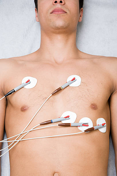
ECG electrodes are thin metal discs that are placed on the patient’s chest, back and arms. The electrodes measure electrical activity in the heart and transmit this information to the ECG machine.
The electrodes are connected by wires to an ECG machine which records the electrical signals from each electrode. This is known as an electrocardiogram (ECG). The ECG machine converts these electrical signals into a form that can be easily interpreted by a doctor or nurse.
The placement of the electrodes on the patient’s body is very important for accurate results. The chest pads are placed directly over the ribs, while the arm electrodes are placed just above the wrist (on top of the forearm).
ECG electrodes are wires that are placed on your chest and back. They are attached to a machine called an electrocardiograph (ECG or EKG) that records the electrical activity of your heart.
Electrodes are small metal disks that stick to your skin using adhesive pads. The electrodes pick up tiny electrical signals from nearby muscle tissue. Some of these signals travel through the heart’s chambers and valves, while others travel through the blood vessels in the lungs and other parts of the body.
An electrocardiograph (ECG or EKG) is a device that records the electrical activity of the heart. It is a noninvasive diagnostic tool that provides an electrocardiogram of the heart over a period of time (24 hours or more).
The acronym ECG stands for “electrocardiograph”. The term “electrocardiogram” is often used as a synonym for ECG, but it is more correctly applied to the recording produced by the machine, whereas an ECG may be recorded by several types of devices.
How many electrodes are on an ECG?
There are 12 electrodes on an ECG. The leads are attached to the 12 electrodes. The electrodes are spread across the chest and arms.
The reason for this is that there are areas of the heart that will be missed if only one lead is used. The 12 leads give us a more complete picture of what is happening in the heart.
The electrocardiogram (ECG or EKG) is a test that measures the electrical activity of the heart over a period of time. It uses electrodes placed on the skin to record the electrical signals produced by the heart.
The number and placement of electrodes varies, depending on what part of the heart you want to see and what type of information you’re trying to collect.
The standard ECG has 12 electrodes in four groups:
1 — two leads attached to two chest electrodes placed on the chest along with one lead attached to an arm electrode (lead I)
2 — three leads attached to three limb electrodes placed on the limbs, one on each leg (leads II, III) and one on each arm (lead aVR).
The ECG electrodes are the most important part of an ECG. They are the reason that we can get a snapshot of the electrical activity in our heart.
The standard ECG configuration has 12 leads, each with 3 electrodes (one positive, one negative, and one ground). The leads are placed on the patient’s skin according to a system developed by Einthoven in 1903.
The ECG electrodes themselves can be either disposable or reusable. Reusable electrodes tend to be more expensive but they last longer and are more comfortable for patients because they can be cleaned between uses. Disposable electrodes have an adhesive backing that sticks to the patient’s skin and must be disposed of after use.
An ECG is a test that records the electrical activity of the heart. It can be used to diagnose heart disease, detect abnormal rhythms (arrhythmias) and assess damage after a heart attack.
The ECG machine has 12 leads. Two are placed on each wrist and two are placed on each ankle. The remaining six are placed over different parts of the chest – some as close as possible to the heart itself, others further away.
Each lead gathers information from a different part of the heart muscle, allowing doctors to see how each part is working independently or together with others
Where ECG electrodes are placed?
ECG electrodes are placed on the chest, arms and legs. These electrodes are used to measure the electrical activity of the heart. They also detect how well your heart is pumping blood around your body.
In this post, we will discuss about where ECG electrodes are placed.
What are ECG Electrodes?
The heart generates electricity that flows through your body in waves. It is measured by attaching electrodes to different parts of the body by adhesive pads or sponges soaked in gel or saline solution. The electrical activity from each electrode is recorded on paper printouts or computer screens as a wave pattern called an electrocardiogram (ECG). An ECG provides information about your heart’s rhythm and structure as well as its electrical activity.
The standard ECG, or electrocardiograph, uses 10 electrodes placed all over the body. The electrodes are connected to an amplifier and the amplified signals are displayed on paper or a computer screen as waves that show various electrical activities in the heart.
The left arm is used for limb leads I and II, which record information about depolarization of the upper left side of the heart. The right arm is used for limb leads III and aVF, which also record information about depolarization of the upper right side of the heart. The chest leads V1 through V6 record information about depolarization of different regions in the lower section of the heart.
The ECG recording is essentially a snapshot of what is happening inside your heart at that moment.
Electrocardiography (ECG or EKG) is a technique for recording the electrical activity of the heart over a period of time using electrodes placed on the skin. The resulting trace is known as an electrocardiogram (ECG). Related techniques that record electrical signals from internal organs include electroencephalography (EEG) for recording brain activity and electromyography (EMG) for recording muscle and nerve activity.
The first practical ECG was invented by Willem Einthoven in 1903 and received the Nobel Prize in medicine in 1924. The invention of the ECG is regarded as one of the most important milestones in medical technology.[1]
The ECG has three leads:
Lead I: The first limb of the circuit
Lead II: The second limb of the circuit
Lead III: The third limb of the circuit
How do you place 3 electrodes on an ECG?
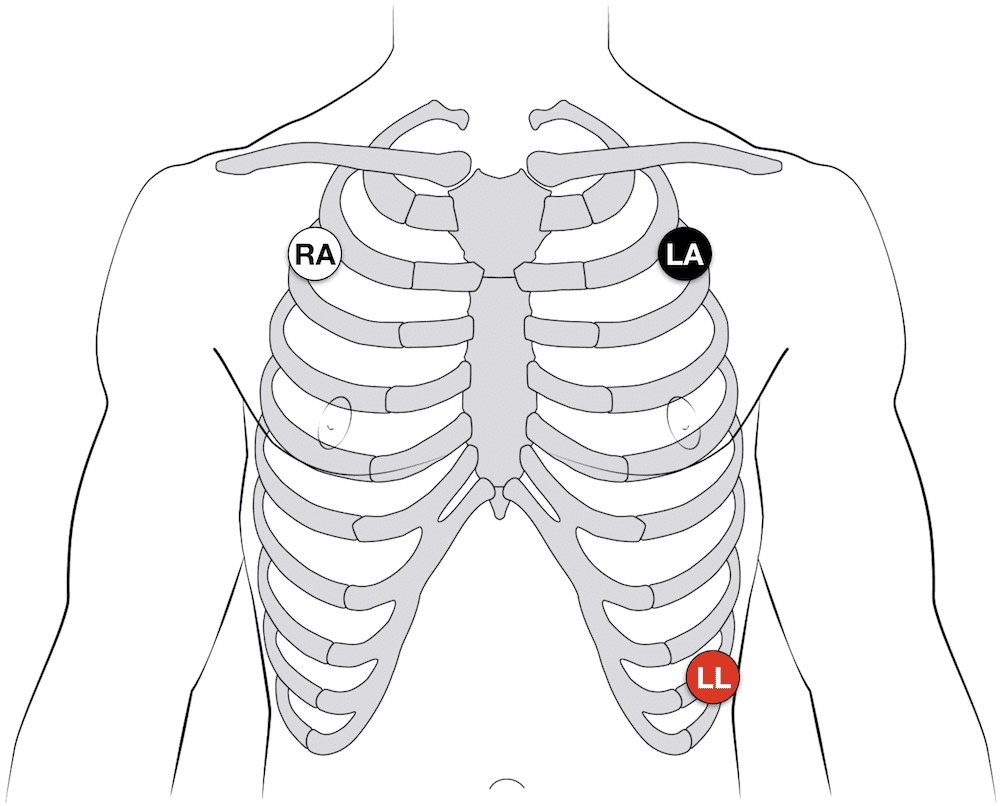
The 3 electrodes are attached to the patient’s chest. The first is attached to the left arm, the second is placed on the right arm and the third is placed on the left leg.
The purpose of ECG is to measure electrical activity of the heart so that any abnormalities can be detected. It is a non-invasive procedure and only takes a few minutes.
The electrocardiogram (ECG) is one of the most important diagnostic tests performed today, especially in cardiac patients. It provides valuable information about how well your heart is working and whether it could be under stress or strain.
The electrodes are usually placed on the chest, one on the right side of the body and two on the left.
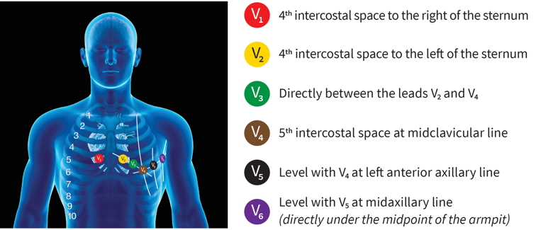
The chest leads are called V1 and V6, which are located at 1 cm above and below the midpoint of a line drawn between nipple line and xiphoid process (see diagram).
The limb leads are positioned on the arms, with a positive electrode at each wrist and negative above or below the wrist.
Lead aVR is simply placed in an appropriate position on the right arm or leg, depending on whether you want to record it as an augmented lead (aVR) or unaltered ECG trace (V5).
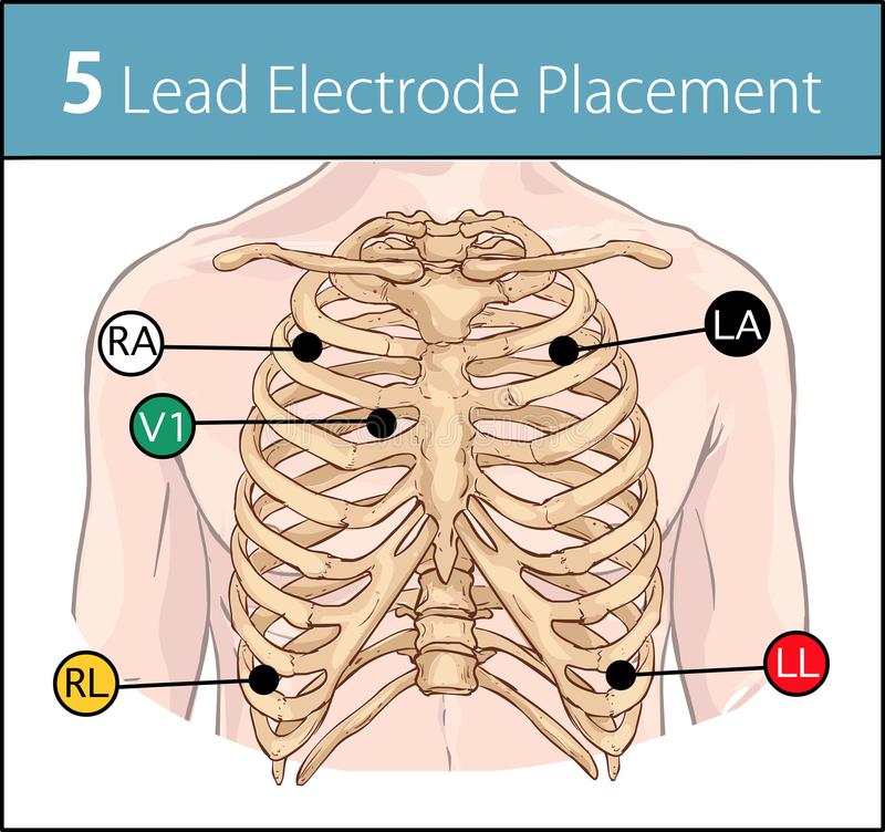
The 10-lead ECG is the most common type of ECG. The 10 electrodes are placed on the chest and limbs, as shown in the following illustration:
There are five limb electrodes, which are placed on each arm and leg.
There are three precordial (chest) electrodes, which are placed near the top of the chest.
The fifth limb electrode is placed below the right shoulder on the 5th intercostal space (ICS).
Each lead records a different electrical signal from a specific part of the heart muscle. The five limb leads record signals from depolarization at different places along the axis of each arm and leg. The three precordial leads record signals from depolarization at different places along the axis of each lung cavity (right superior pulmonary vein, left superior pulmonary vein, posterior wall).
The placement of electrodes on the body is a matter of convenience and personal preference. However, there are some basic rules that should be followed.
Lead I (the red electrode) is usually placed on the right arm or chest. Lead II (the green electrode) is placed at the left side of the chest or on the left arm. Lead III (the white electrode) is placed on the right leg.
The location of leads I, II and III determines what information will be recorded by each lead.
What are the 3 types of ECG?
There are three types of ECG:
Normal (sinus rhythm): Normal heart rate is between 60 and 100 beats per minute. The QRS complex measures 0.06 to 0.10 seconds, and the P wave follows the QRS complex by 0.04 seconds or less.
Atrial fibrillation: This condition causes the atria to beat too quickly, resulting in a disorganized, irregular heartbeat that can be hard to detect on an ECG. However, it’s important to get this condition treated because it can lead to blood clots and stroke.
Ventricular tachycardia: This is a dangerous arrhythmia that occurs when one of your ventricles (lower chambers) beats too quickly — usually more than 150 times per minute.
There are three types of electrocardiogram (ECG):
12-lead ECG. This is the most common type of ECG. It measures electrical activity from 12 different leads on the body.
24-lead ECG. This test records electrical activity from 24 different leads on the body. It gives more information about electrical activity than a 12-lead ECG and can help detect more problems with heart rhythm and heart muscle contraction than a 12-lead ECG can.
Holter monitor. This is a portable device that you wear while at home or at work for 24 to 48 hours or longer to record your heart’s electrical activity continuously throughout this time period. A doctor may recommend this test if you have symptoms like palpitations or dizziness that might be caused by abnormal heart rhythms
There are three types of ECG:
The standard 12-lead ECG, which is the most common type of ECG. The standard 12-lead ECG is used to measure and monitor the heart rate and rhythm (arrhythmias), as well as the electrical activity within the heart. The 12 leads measure the electrical signals that pass through your heart from one side to another, and record it onto a graph paper called an electrocardiogram (ECG).
The resting ECG, which measures your heart’s electrical activity while you are at rest. This test is usually done first thing in the morning after you have been lying flat for 10 minutes or more. This test can be useful for diagnosing certain conditions that cause an abnormal heart rate or rhythm.
The stress ECG, which measures your heart’s electrical activity during exercise or other stressors. This test can be done with an exercise machine or by walking on a treadmill while wearing a portable device connected to electrodes placed on your chest.
What is 5 lead ECG used for?
ECG is the abbreviation for electrocardiogram, which is a graphical representation of the electrical activity of the heart. An ECG records the electrical potentials generated by the heart and the flow of these potentials through the heart muscle. The ECG can be used to detect abnormal rhythms and to assess cardiac function.
5 lead ECG is a standard ECG that records five main leads: V1, V2, V3, V4 and V5. These are arranged in an equilateral triangle pattern with each lead representing a different view of the heart. In other words, there are three views (V1-V3) from above and two views (V4-V5) from below.
Electrodes are connected to each lead so that when an action potential travels through the heart, it will produce a small voltage drop at each electrode’s location on the chest wall. These voltages are then amplified and recorded on paper or digitally as an electrocardiogram (ECG).
5 lead ECG is a type of ECG that can be performed on patients who are unable to lie down. It is also a way to detect arrhythmias in patients who have difficulty lying flat on their backs.
The 5 leads used are the V1, V2, V3, V4, and V5 locations. These leads are placed on the chest in a similar fashion as 12-lead ECGs. The placement of these leads is not as precise as 12-lead ECGs because they do not need to be placed in exactly the same spot every time.
The lead placement for 5-lead ECGs may vary depending on whether or not you are using an automatic machine or manual machine. The only difference between the machines is that automatic machines will make all of the necessary calculations while manual machines require you to add up all of the voltage values yourself before recording them on paper or digitally (e.g., on a computer).
Why is a 12 lead called a 12 lead?
It’s called a 12-lead because it has 12 electrodes, one for each of the 12 main arteries that supply blood to the heart.
Each electrode is attached to a wire that connects it to the heart monitor. The wires connect to the monitor through two sticky pads that are placed on your chest. When you lie down, these pads hold the wires in place so that they don’t move around as much as they would if you were sitting or standing up.
The wires are connected to a small disc that you place on your upper back. This disc contains all of the electronics needed to convert your heart’s electrical signals into numbers and waveforms that can be read by a doctor or nurse in real time at their computer screen.
The doctor or nurse can use this information to determine if there are any abnormalities in your heart’s electrical activity (the electrocardiogram (ECG), which is also known as an EKG).
A 12-lead ECG is a set of 12 simultaneous recordings of the electrical activity of the heart. It is obtained by placing electrodes on the body, typically on the chest and limbs, and sometimes on other locations, such as the neck.
The data from each electrode is sent to a 12-lead display unit that shows all 12 leads at once.
The leads are labeled I through XII, with lead I being closest to the heart in between the left arm (LA) and left leg (LL), and lead IX being closest to the heart in between the right arm (RA) and right leg (RL). A normal heart has an electrical potential difference between its right and left sides; however, there are several types of abnormalities which can occur due to various types of heart diseases. These abnormalities show up as changes in voltage or current patterns on an ECG tracing.