An ultrasound of pyloric stenosis is a medical test that uses sound waves to create pictures, or images, of the structures inside your body. The test is done to look at your child’s stomach and see if there are any problems with it.
The ultrasound uses a small wand called a transducer. A gel is placed on the area to be examined. The gel helps transmit the sound waves. The transducer sends sound waves into your child’s body and picks up the echoes as they bounce off the organs inside. A computer uses the echoes to create an image of the tissues and organs inside your child’s body.
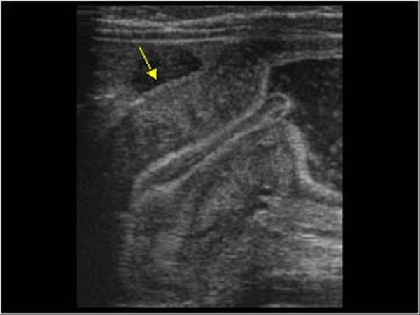
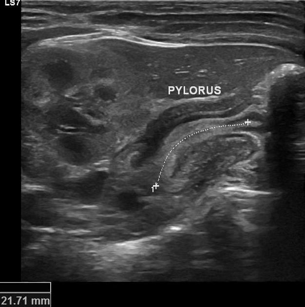
The ultrasound also shows pyloric muscle thickness of more than 4 mm, a criterion that has been shown to have a sensitivity of 96% and a specificity of 92%.
The treatment for pyloric stenosis is surgery. In the past, this was performed with an open surgical procedure. More recently, pyloromyotomy is performed laparoscopically. The results are excellent and complications are minimal.
The diagnosis of pyloric stenosis is usually made clinically by observing the projectile vomiting and feeling the olive-shaped mass palpated in the epigastrium. The presence of nonbilious, projectile vomiting should prompt an abdominal radiograph to rule out malrotation and midgut volvulus when there is associated bilious vomiting or abdominal pain. Ultrasonography is useful in confirming the diagnosis and ruling out other causes of vomiting such as intussusception.
Pyloric stenosis is a condition that affects infants in which the opening between the stomach and the small intestine (called the pylorus) becomes narrowed. With pyloric stenosis, food cannot leave the stomach and builds up, causing vomiting. As a result, infants lose weight and become dehydrated.
Ultrasound can help diagnose pyloric stenosis in some cases by showing thickening of the wall of the pylorus or blockage of movement of food through it. Many other conditions can cause vomiting in infants, however, so additional tests may be needed to confirm a diagnosis of pyloric stenosis.
Pyloric stenosis is a stenosis (narrowing) of the pylorus. The pylorus is the part of the stomach that connects to the duodenum. The narrowing prevents food from emptying from the stomach into the small intestine. Pyloric stenosis is most common in infants, especially males. If not treated, vomiting will occur after eating and weight loss or dehydration may result.
Pyloric stenosis can most often be diagnosed based on the symptoms. Ultrasound imaging may confirm the diagnosis.
Treatment involves surgery to widen the pylorus. Without treatment, complications may include pneumonia and malnutrition.[1] The condition affects about 3 in every 1,000 live births.[4] When it does occur, it most commonly begins between two and eight weeks of age.[1]
What is pyloric stenosis?
Pyloric stenosis is a problem with the valve (pylorus) between the stomach and small intestine that allows food to pass into the small intestine. Normally, when you eat, food goes through your esophagus into your stomach. The pyloric sphincter regulates the flow of food out of your stomach. In pyloric stenosis, the valve connecting your stomach and small intestine becomes blocked by tissue. This blocks the flow of food from your stomach to your intestines.
Pyloric stenosis is more common in male than female babies. It can also occur in older children and adults.
An enlarged pyloric canal (normally < 12 mm in length) with incomplete relaxation of the pyloric sphincter, resulting in gastric outlet obstruction.
It is due to hypertrophy and elongation of the muscle fibers of the pyloric region. It can be congenital or acquired (eg, as a result of infection, inflammation, or trauma)
The Gastrointestinal Tract
The gastrointestinal (GI) tract consists of the esophagus, stomach, small intestine, and large intestine. The GI tract is responsible for digesting food, absorbing nutrients, and eliminating waste.
Ultrasound is a type of imaging test that uses sound waves to create pictures of structures inside the body. Ultrasound is used to examine many parts of the body including:
Abdomen
Pelvis
Thyroid gland
Eye sockets
Blood vessels
How Does An Ultrasound Detect Pyloric Stenosis?
An ultrasound is typically used to detect pyloric stenosis. This is a non-invasive imaging technique that doesn’t involve the use of radiation. An ultrasound can be used to visualize the pylorus and determine if it has narrowed.
The pylorus is located at the bottom of the stomach, where it meets with the first part of the small intestine (the duodenum). The pyloric sphincter is a circular band of muscle that sits around this opening. When it contracts and relaxes in coordination with peristalsis, food can pass into the duodenum.
During an ultrasound examination, sound waves are sent into the body and bounced back by different structures. These signals are received by a computer, which uses them to generate an image of the baby’s internal organs on a monitor.
Ultrasounds are considered safe for both mother and baby. There are no known harmful effects from exposure to sound waves at this frequency. Although an ultrasound is not completely risk-free, it is much safer than X-ray imaging when done correctly
An ultrasound is a noninvasive imaging test that uses sound waves to produce pictures of what is inside your abdomen. It is used to visualize the internal organs and structures of the abdomen. This test can be used to diagnose pyloric stenosis, as it can show the thickening of the pylorus muscle (the opening between the stomach and small intestine).
Ultrasound is a very useful diagnostic tool for pyloric stenosis. It has high sensitivity and specificity in detecting pyloric stenosis in neonates. It is a noninvasive and painless diagnostic test that can be repeated multiple times without any harm to the patient. Ultrasound findings include thickened pylorus greater than 4 mm, elongated pylorus length more than 14 mm, and non-visualization of the air bubble in the stomach. Transient elastography is also an upcoming technique for the diagnosis of pyloric stenosis, as it can detect muscle thickness with good accuracy.
Pyloric stenosis is a condition of the stomach that is congenital (present from birth) but not often diagnosed until a few weeks after birth. This disorder affects the stomach and the pylorus, which is the passage between the stomach and the small intestine. In some babies, this passageway (pyloric canal) enlarges abnormally, making the exit of food from the stomach difficult.
Echocardiogram: An echocardiogram uses sound waves to produce detailed images of your heart’s chambers, valves and walls. An echocardiogram can detect heart disease, such as damaged or thickened heart muscle, problems with your heart’s valves or problems with blood flow through your heart.
The pylorus, or pyloric canal, is a small passage that connects the stomach to the duodenum. The stomach produces an enzyme called pepsin, which helps break down food. Enzymes from the pancreas and bile from the liver also aid in digestion. These enzymes are added to the chyme as it moves from the stomach into the duodenum.
Pyloric stenosis occurs when the pylorus becomes inflamed and thickens, causing it to narrow. This prevents food from moving through as quickly as it should. When this happens, stomach acid can back up into your baby’s throat, causing vomiting.
An ultrasound test is a procedure that uses high-frequency sound waves to scan a woman’s abdomen creating a picture (sonogram) of the baby and placenta. Ultrasound exams do not use ionizing radiation (as used in x-rays). Because ultrasound images are captured in real-time, they can show the structure and movement of the body’s internal organs, as well as blood flowing through blood vessels.
Ultrasound imaging is a noninvasive medical test that helps physicians diagnose and treat medical conditions.
Ultrasound exams do not use radiation (as used in x-rays). Because ultrasound images are captured in real-time, they can show the structure and movement of the body’s internal organs, as well as blood flowing through blood vessels. Ultrasound imaging is often used to evaluate symptoms such as pain, swelling and infection that may be due to complications with organs, tissues or abdominal structures within a woman’s pelvis.
Ultrasound uses sound waves to create images of structures within your body. In an ultrasound, a technician spreads a warm gel over your abdomen and moves a device called a transducer across your skin. The gel helps the transducer pick up images from inside your body.
Ultrasound is often used to guide procedures, such as needle biopsies, in which a sample of tissue is extracted for testing.
Ultrasound imaging doesn’t involve radiation. It’s usually painless, though it may be uncomfortable if you have to hold a position for long periods of time or if the technician presses hard on your abdomen to get better images.
Ultrasound isn’t recommended during the first trimester of pregnancy because the developing baby may be harmed by high frequency sound waves.
What is The Normal Measurement For Pylorus?

What is the normal measurement for pylorus?
In the adult, the pyloric canal is about 2 to 3 cm long and is lined with a layer of circular muscle. The mucosa of the pyloric antrum has mucous glands, which produce mucus. The mucosa of the pyloric canal contains numerous gastric pits and few mucous glands.
Is this answer helpful?
What is the normal measurement for pylorus?
INTRODUCTION: The pylorus is a muscular sphincter that connects the stomach to the duodenum. It prevents the reflux of gastric contents into the duodenum and regulates the passage of food from the stomach to the duodenum.
We hypothesized that the pylorus length would vary according to age, gender, and body mass index (BMI).
The measurement of the pylorus is usually measured at about 2 to 4 cm in adults and 1.9 to 3.4 cm in children.
The pylorus is the lower portion of the stomach that connects to the duodenum. This part of the stomach is responsible for keeping food in the stomach until it is ready to be digested. The pylorus also regulates the flow of food from the stomach into the intestine.
The pyloric sphincter is a muscular ring that regulates this process by relaxing and contracting to allow food to leave the stomach at a controlled rate.
Pyloric stenosis occurs when this opening becomes so narrow that food cannot pass through it or can only pass through slowly, causing the stomach to become enlarged.
Pylorus measurements are not commonly used in medicine, but may be done if your doctor suspects you have a pyloric disorder such as pyloric stenosis or gastric outlet obstruction.
It is difficult to give a single measurement for the pyloric canal. There are both vertical and horizontal measurements that can be taken.
The horizontal measurement runs from the pylorus to the antrum and is approximately 2.5 cm long.
The vertical measurement is a bit more complicated. The muscle of the pylorus is sphincter-like in nature and can be everted and inverted by applying a force to it. When relaxed, it has an average length of 1.9 cm but can stretch to 3 cm when everted. Similarly, it can be inverted to a length of 0.7 cm on average, but can reach down to 0.3 cm in length when fully inverted (this is likely due to the presence of mucosa covering the inner aspect of the pyloric muscle).
Pyloric stenosis is a condition in which the pylorus – the opening from the stomach into the small intestine – becomes narrowed, causing food to move slowly or stop moving into the small intestine. This causes vomiting after eating. Pyloric stenosis usually occurs within the first few weeks of life.
The pylorus is a 1-2 cm long muscular tube joining the stomach to the duodenum (the upper part of the small intestine). The pylorus controls how fast food moves from your stomach into your intestines.
Pyloric stenosis usually develops before birth, when the infant’s pylorus muscles grow larger than normal and become thickened. However, it’s often not diagnosed until later in infancy.
The cause of pyloric stenosis isn’t always clear. It may be linked to genetics, as it often runs in families. Pyloric stenosis is also more common among firstborn infants and twins, as well as males and babies who are born to mothers older than 35 years of age.
Symptoms of pyloric stenosis include:
vomiting immediately after feeding
vomiting that contains undigested food hours after feeding
dehydration due to loss of fluids through vomiting

The pylorus, or pyloric antrum, is the lower portion of the stomach that connects to the duodenum (the first segment of the small intestine). Its purpose is to release chyme into the duodenum. The pylorus is surrounded by a thick layer of smooth muscle which controls the release of food from the stomach.
The pylorus has three functions:
It stores food coming from the esophagus until it can be emptied into the duodenum.
It controls the flow of food into the duodenum by regulating gastric motility and emptying.
It mixes gastric contents by contracting in a rhythmic fashion.