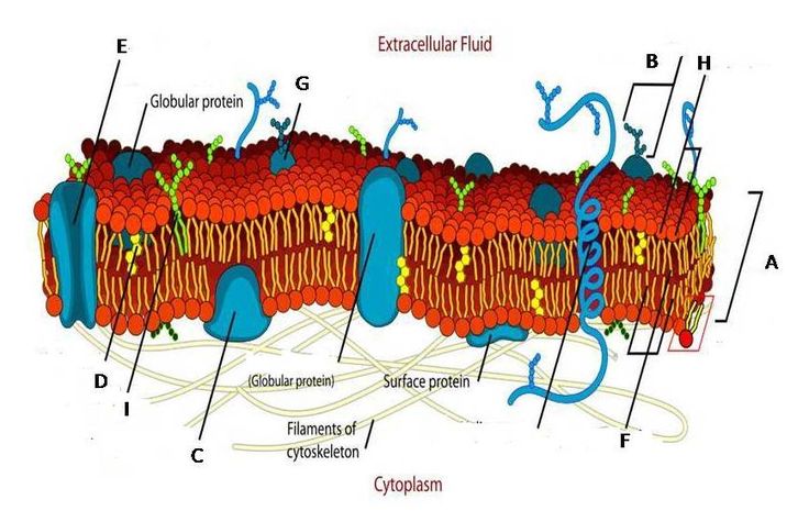
Label the Structures Of The Plasma Membrane and Cytoskeleton.; The plasma membrane is the outermost structure of a cell, the cellular boundary between what is inside the cell and what lies outside. The glycoproteins that form the exterior surface of all cells are important in cell-cell recognition and interactions, as well as in interacting with extracellular materials. Glycolipids with their glycerol heads also form part of the membrane surface. Glycoproteins are important in attaching to other extracellular materials, such as collagen fibers and components of basement membranes.
Intrinsic proteins can be found embedded within the lipid bilayer forming channels through which ions and molecules can pass, or they can span across both sides of the membrane acting as structural components of enzymes. In addition, some integral proteins have carbohydrate chains attached to them which protrude out from the surface of the cell, forming glycoproteins (glyco means sugar).
Peripheral proteins are loosely attached to either side of the cell membrane by ionic bonds or hydrogen bonds. These proteins are often enzymes that catalyze various reactions at or near the cell surface.
In most mammalian cells, cholesterol is an abundant constituent of the plasma membrane. Cholesterol intersperses itself among
The plasma membrane (also known as the cell membrane) is a thin semi-permeable membrane that surrounds the cytoplasm of a cell. Its function is to protect the integrity of the interior of the cell by allowing certain substances into the cell while keeping other substances out. It also serves as a base for chemical signals to pass in and out of the cell.
The structure of the plasma membrane is very similar to a lipid bilayer. The plasma membrane contains phospholipids, cholesterol, proteins and carbohydrates. The phospholipids form two layers with hydrophobic tails facing inward and hydrophilic heads facing outward.
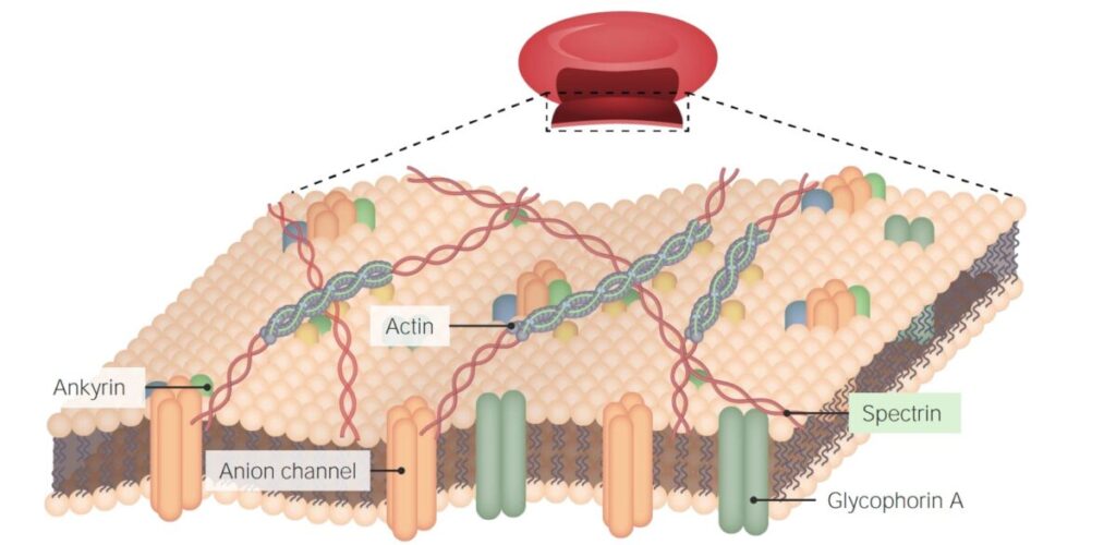
The cytoskeleton is a network of fibers composed of three types of protein filaments: microtubules, microfilaments, and intermediate filaments. These fibers provide support and structure to the cells they inhabit. They also give cells their shape, enable movement, and allow cells to move other objects around inside them.
Plasma Membrane
The plasma membrane is the outer boundary of a cell. It is semi-permeable and allows some substances to enter and exit the cell. The plasma membrane also contains proteins that identify the cell as part of a particular organism.
Cytoskeleton
The cytoskeleton maintains the shape of the cell, provides structure for organelles, anchors organelles in place, and allows for movement within the cell. The cytoskeleton is made of three main structures: microtubules, microfilaments, and intermediate filaments.
The plasma membrane is the outermost boundary of a cell. It is a dynamic structure, constantly changing in composition and shape. Because it separates the cells from the external environment, it regulates what enters and leaves the cell.
The plasma membrane consists of two layers of phospholipids – also called a phospholipid bilayer – with proteins embedded in it. This model of cell membrane is known as the fluid mosaic model because lipids and proteins can move within the membrane (like a mosaic) and because lipids move around easily (like a fluid). As shown in Figure 1, the phospholipid heads form an inner “sea” of hydrophilic (water-loving) molecules, whereas their hydrophobic (water-fearing) tails are oriented away from this sea towards the surrounding environment. An important property of this arrangement is that phospholipids cannot mix with water; they will spontaneously form bilayers to separate themselves from water.
The fluid mosaic model also describes how membrane proteins are distributed within the plasma membrane. Two major types of events are driven by these proteins: endocytosis and exocytosis. Endocytosis refers to movement of molecules into the cell, while exocytosis refers to movement out of the
- The cytoskeleton is a network of protein filaments, microtubules and actin filaments that support the cell and give it shape. In addition, it plays an important role in cell movement.
- Actin filaments are made from the protein actin. The actin filament is composed of two strands of globular actin that twist around each other to form a helix. A protein called tropomyosin lines the groove created by the helix and prevents myosin from binding to actin.
- Intermediate filaments are composed of a variety of proteins, including those found in desmosomes, keratin, and nuclear lamina.
- Microtubules are made from tubulin subunits that assemble into long tubes with 13 protofilaments on the inside and 14 protofilaments on the outside, giving them a diameter of 25 nanometers (nm). Microtubules are also called hollow cylinders because they are tubular in shape with a hollow center, like a pipe or straw. Microtubules play an important role in cell structure and function by providing support for the cell and a mechanism for moving materials within it.
What is The Structure of Plasma Membrane?

The plasma membrane is a bilayer of lipid molecules with many proteins embedded in it.
The phospholipid bilayer (see diagram on the right) is a thin polar membrane made of two layers of lipid molecules. These lipids have hydrophilic (water-soluble) “heads” and hydrophobic (water-insoluble) “tails”. The hydrophilic heads face outwards and are next to the water in the cytoplasm or the extracellular fluid. The hydrophobic tails face each other, in the middle of the membrane.
The molecular organization of the membrane gives it different properties. For example, it is permeable to small, uncharged molecules like oxygen and carbon dioxide but not to large, charged molecules like proteins and nucleic acids. This property is essential to life: it allows cells to regulate what goes into and out of the cell.
The plasma membrane is a phospholipid bilayer in which proteins are embedded.
The proteins may be inserted into the lipid bilayer or attached to the surface of one of the phospholipids layers.
The proteins can also be attached to carbohydrates, which are on the outside of the plasma membrane or anchored to other proteins within the phospholipid bilayer that are extending through it.
There are two types of proteins: peripheral and integral.
The plasma membrane is a thin and structured bilayer of phospholipid and protein molecules that surrounds the cytoplasm of a cell. It contains many different types of proteins embedded in the phospholipid bilayer or attached to the outer surface of the membrane. These proteins perform various functions, including transport of substances across the membrane, cell-to-cell recognition, enzyme activity, attachment to the cytoskeleton and extracellular matrix, etc. The phospholipids in the plasma membrane are asymmetrical; that is, they consist of two different types of lipid molecules. The “head” region of each lipid molecule is hydrophilic (water-loving), while the “tail” region is hydrophobic (water-hating). All these polar head groups are oriented towards the aqueous environment outside and inside the cell; all nonpolar tails are oriented inwards towards each other, away from any water. This type of structure creates a fluid mosaic model for membranes with embedded proteins.
A unique feature of the plasma membrane is that it is a semi-permeable barrier. This means that it allows some substances to pass across it more easily than others which require an active transport mechanism. The membrane is composed of a phospholipid bilayer with protein molecules embedded in it.
Phospholipids are compounds that consist of a phosphate group, two fatty acids and glycerol. The fat soluble hydrophobic ends of the phospholipids aggregate together leaving the hydrophilic phosphate groups on the outside forming a barrier to hydrophilic (water soluble) materials.
The proteins perform a number of functions including providing cell identity with surface antigens, providing attachment sites for extracellular structures like the cytoskeleton and extracellular matrix, acting as receptors for hormones and growth factors and providing transport mechanisms for hydrophilic molecules.
The plasma membrane is made up of a phospholipids bilayer with proteins and glycoproteins embedded in it.
Phospholipids are arranged so that their hydrophobic tails are pointing toward each other within the membrane.
Proteins form channels that allow substances to move through the membrane, while others act as receptors that bind to hormones or molecules so cells can respond to them.
Glycoproteins have carbohydrates attached to them and function as cell markers that help other cells recognize the cell that bears these proteins.
For example, T cells (white blood cells) will look for glycoproteins on a virus-infected cell and destroy it.
The plasma membrane is a double layer of phospholipids, with embedded proteins. Phospholipids have a polar (hydrophilic) head and two non-polar (hydrophobic) tails. The polar heads are on the outside and the hydrophobic tails are on the inside. The phospholipids are arranged so that the heads form a barrier between the cell’s contents and its surroundings.
Phospholipid molecules are asymmetrical; they have a “head” which is water-loving (hydrophilic), and two “tails” which are water-hating (hydrophobic). Phospholipid molecules spontaneously arrange themselves into a bilayer structure, so that their polar heads point outwards, towards the watery environment both inside and outside of the cell, while their non-polar tails point inwards, away from the watery environment. This arrangement forms a boundary between the cytoplasm inside the cell, and its surroundings outside of the cell.
The plasma membrane is the outermost layer of the cell and is composed of a phospholipid bilayer. Embedded in the bilayer are proteins, which perform important functions for the cell, as well as carbohydrates that function as identifiers.
Phospholipids (water-fearing)
Is the Plasma Membrane a Part of The Cytoskeleton?
No, the plasma membrane is not a part of the cytoskeleton.
The cytoskeleton is composed of microtubules, actin filaments, and intermediate filaments that are made out of protein. The plasma membrane however is composed of cholesterol and phospholipids.
The plasma membrane is not a part of the cytoskeleton. Instead, it’s the boundary between the cell and the extracellular environment. It regulates the communication between the cell and its environment.
The plasma membrane is not part of the cytoskeleton (which includes actin filaments, microtubules and intermediate filaments). The plasma membrane is a phospholipid bilayer embedded with proteins that forms the outer boundary of a cell. The cytoskeleton includes filamentous scaffolds that help to give the cell its shape, provide mechanical stability and allow for organelle movement, but does not include the membranes of the cell.
In animal cells, the plasma membrane is not part of the cytoskeleton. But it interacts with the cytoskeleton in several ways:
The cytoskeleton provides mechanical support to the plasma membrane by embedding it in a network of actin filaments and microtubules. This helps keep the cell spherical and keeps its shape stable.
The cytoskeleton plays a role in transporting materials within the cell. It’s possible that some of these transport mechanisms involve both microtubules and actin filaments — for example, vesicles may use kinesin to move along microtubules, while at the same time being anchored to actin filaments by myosin motors.
Cytokinesis involves the assembly of an actomyosin contractile ring around the plasma membrane which contracts and pinches off the daughter cells from each other.
No. The plasma membrane is the outer boundary of a cell. The cytoskeleton is inside the plasma membrane, in the interior (cytoplasm) of a cell.
The cytoskeleton is a network of fibers that run throughout the cell. It is composed of three types of protein filaments:
Microfilaments (actin filaments) – 7 nm in diameter, these are involved in muscle contraction, amoeboid movement of cells, and cytokinesis. They are made up of actin molecules. Microfilaments also play a role in the motility (movement) of cilia and flagella.
Microtubules – 25 nm in diameter, these form the “skeleton” of cilia and flagella, which are composed of bundles of microtubules called axonemes. They are made up of tubulin molecules.
Intermediate filaments – 10 nm in diameter, these provide structural support to the cell as well as anchorage for organelles. They are made up of various proteins such as keratins and neurofilaments.
The plasma membrane is not part of the cytoskeleton because it is a lipid bilayer arranged into a sheet that surrounds the entire cell, whereas cytoskeletal elements run throughout the entire cell.
The plasma membrane is the cell membrane of a cell. It is a selectively-permeable barrier that allows some substances to cross it more easily than others. The permeability of the plasma membrane allows nutrients and wastes to move in and out of cells, while at the same time preventing larger molecules from getting into or out of cells.
The cytoskeleton is an internal network of protein filaments within cells. It acts like a “skeleton” for eukaryotic cells, providing support for the cell, as well as enabling it to change shape and move. The cytoskeleton is made up of three types of protein filaments: microfilaments (actin filaments), intermediate filaments, and microtubules.
The plasma membrane does not have the same role as that of the cytoskeleton. Instead, it functions as a selectively-permeable barrier between the cell’s internal environment and its extracellular environment. It also plays roles in cell signaling, as well as in endocytosis and exocytosis.