Podocytes Kidney; podocyte nucleus and cytoplasm is usually rich in ribosomes. The cytoplasm contains numerous fibrils, which due to the presence of actin filaments are contractile.
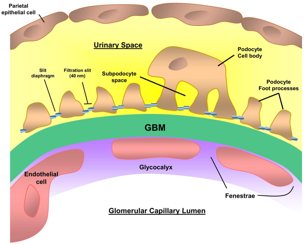
Podocytes are specialized visceral epithelial cells. In the kidney, they cover the surface of the glomerular capillaries, a function analogous to the squamous epithelial cells covering the non-glandular portion of the stomach. Podocytes have foot processes, known as pedicels or “feet”, that interdigitate with one another creating a filtration barrier to prevent large molecules from passing through. This forms a barrier using slits, termed filtration slits, between adjacent foot processes (also called pedicels). These slits are formed by podocyte cell membrane proteins nephrin, CD2AP and podocin. The slit pores between two podocyte cells are approximately 30-40 nanometers wide.[1]
Loss of this filtration barrier leads to proteinuria and thus kidney disease. In addition to their filtering function, podocytes also possess endocytic activity as they are able to internalize albumin via clathrin-mediated endocytosis.[2]
Podocytes are cells that make up the filtration unit of the kidney, and when they become damaged, protein leaks into the urine.
The kidney is made up of millions of small filtering units called nephrons. Each nephron filters waste products from the blood into the urine. These waste products include excess water, salts and urea, a nitrogen-containing waste product from protein metabolism.
Nephrons contain two parts:
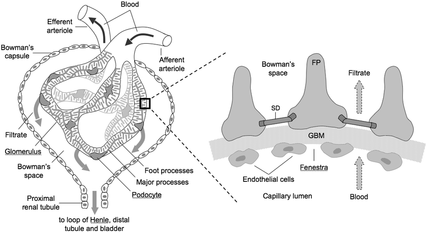
Glomerulus — a cluster of capillaries (tiny blood vessels) surrounded by a capsule called Bowman’s capsule that collects waste products from the blood
Bowman’s capsule — surrounds the glomerulus; made up of two layers: an outer layer of simple squamous epithelial cells and an inner layer of podocytes (visceral epithelial cells)
Podocytes are cells that wrap around the capillaries in your kidneys. They have projections known as foot processes that reach into the capillaries and filter out wastes from your blood. In nephrotic syndrome, these foot processes are damaged, which prevents the podocytes from functioning normally.
When podocytes are damaged, some waste products can no longer be filtered from your blood, leading to proteinuria (protein in the urine), hypoalbuminemia (low albumin levels in the blood), and edema (water retention).
podocytes are a type of epithelial cell that are located in the filtration slits between adjacent visceral epithelial cells of the glomerulus, a specialized structure in the kidney that filters blood to form urine. Podocytes wrap around capillaries of the glomerulus and branch extensively to form a tuft that contains several hundred foot processes, which play an important role in forming urine. Podocytes are not true cells but shared cells for when they replicate there DNA is not replicated so it is impossible for them to divide.
Podocytes are a type of kidney cell, with foot-like processes called pedicels. They are found in the walls of Bowman’s capsule, which surrounds the glomerulus of the nephron, where blood is filtered to form urine. Podocytes maintain the filtration barrier between blood and the nephron.
Podocytes are characterized by their highly ramified appearance (having many branches). The cell body contains a highly coiled nucleus, and its cytoplasm is abundant in mitochondria, which provide energy for ion transport across its extensive cell surface membrane. The interdigitating pedicels extend from the cell body down into the space between adjacent cells, and are covered with a dense coat of specialized actin filaments called foot processes (or pedicels).
In the mammalian kidney, the podocyte is a highly specialized epithelial cell located at the visceral layer of Bowman’s capsule. Podocytes are characterized by many long foot processes containing slit diaphragms that interdigitate with the foot processes of neighboring cells. The slit diaphragms filter blood through the glomerular capillaries.
The podocyte was first described by German histologist Max Schultze in 1858, who named it “Malpighian body” after Marcello Malpighi, an Italian physician and biologist. In 1911, French pathologist Étienne Jules Marey renamed the structure “podocyte”, from the Greek root pod-, meaning “foot”. However, some authors still refer to this structure as “Malpighian body”.
The renal corpuscle (glomerulus) consists of a glomerular capillary tuft surrounded by a double-layered capsule, the outer layer being continuous with the epithelium of the proximal convoluted tubule and the inner layer consisting of modified visceral epithelial cells (podocytes).
What is The Function of Podocytes Kidney?
Podocytes are the major filtration cells of the kidney. They are important in the formation of urine by filtering blood plasma into Bowman’s capsule. Podocytes have a foot-like structure that wraps around capillaries in the kidneys. They are supported by the glomerular basement membrane, which is composed of laminin and collagen IV.
Podocytes are covered with slit diaphragm proteins, which form pores or slits in between adjacent podocyte cells.
The filtration process occurs when blood pressure forces plasma through these pores into Bowman’s capsule. This filtration is essential to remove toxins and other waste products from blood and make urine.
Podocytes are epithelial cells located at the Bowman’s capsule of the kidney. They have a function of filtration. Podocytes have a huge surface area due to the presence of primary and secondary processes. The primary processes wrap around glomerular capillaries, while the secondary processes are in contact with the lumen of Bowman’s capsule. The podocytes have foot processes that form filtration slits between adjacent cells. This filtering apparatus allows selective passage of water and small molecules into Bowman’s space, which is then excreted as urine. Podocytes are connected by junctional complexes such as adherens junctions, tight junctions, gap junctions and desmosomes, which maintain the integrity of the filtration slits between the cells. If these podocyte-podocyte attachments are disturbed, this can lead to proteinuria (protein in urine). The condition where there is loss or reduction of podocytes from glomeruli is called focal segmental glomerulosclerosis (FSGS). This can be due to genetic mutations (e.g. WT1 gene) or autoimmune disease (e.g. lupus nephritis).
Podocytes are specialized epithelial cells that cover the Bowman’s capsule of each nephron in the kidney.
The primary function of podocytes is to filter blood and produce urine, hence their nickname: “the foot cells.”
The capillary network where the podocytes are located is known as the glomerular capillary network. The glomerular capillary network, made primarily of fenestrated endothelial cells, serves as a filtration barrier.
Podocyte foot processes and slit diaphragms are essential for maintaining the glomerular filtration barrier.
In addition to its role in filtration, the podocyte also plays a role in maintaining the structural integrity of the glomerulus through cell-cell adhesion, cell-matrix adhesion, and protein synthesis.
They are cells in the kidney responsible for filtering fluid from the blood.
Podocytes are specialized epithelial cells that form part of the filtration barrier of the glomerulus. They have a highly complex shape, with long foot processes that interdigitate with those of neighboring podocytes. They are anchored to the underlying basement membrane by integrins, and to each other by adhesion molecules such as connexins.
The podocyte cell body is connected to the glomerular basement membrane by pedicels called foot processes, which are folded extensively and give rise to a large surface area for filtration. The key anatomical features of podocytes include:
podocyte foot processes (primary process), which project into Bowman’s space;
podocyte secondary processes, which are located within Bowman’s space;
pedicules, which connect podocyte foot processes to Bowman’s capsule; and
slits between adjacent pedicles, also known as slit pores.
Podocytes are cells that line the capillaries of the glomerulus (a part of the kidney). They have a very important function – they “filter” out unwanted proteins from the blood and keep them in the blood.
Podocytes get their name because they consist of many small extracellular processes called “pedicels.” Podocytes are very similar to endothelial cells lining blood capillaries, or epithelial cells lining the alveolar sacs (parts of lungs).
The glomerular filtration barrier consists of three layers: fenestrated capillary endothelium, glomerular basement membrane and podocytes. Fenestrated capillary endothelium allows for even large proteins to pass into the glomerulus. Glomerular basement membrane is a thick meshwork of proteins that prevents large proteins from getting back into the vasculature (the system of veins and arteries that carries blood) but allows small molecules such as water and sodium to freely move across. Podocytes are covered with pedicels that form numerous slit pores through which molecules can pass. The slits get narrower and further apart with increasing molecular size, so larger proteins cannot pass through this layer.
The glomerulus is a complex network of capillaries surrounded by specialized cells. Glomerular filtration requires that podocytes cover the glomerular capillaries and maintain a specialized barrier, the glomerular filtration barrier (GFB). The GFB selectively filters molecules from blood into Bowman’s space, a narrow cavity between the podocyte foot processes that is continuous with the lumen of the urinary space.
The GFB includes three cell types: endothelial cells, podocytes and parietal epithelial cells. Each cell type contributes to GFB function. Podocytes must be attached to both the endothelium and parietal epithelium in order to prevent fluid or protein leakage into the urinary space and to allow fluid and protein-free plasma to filter into Bowman’s space.
Podocytes are specialized epithelial cells found on the outer surface of glomerular capillaries. These cells have long foot processes that surround the capillary loops and form slit diaphragms, which are gaps between adjacent foot processes with unique structures that allow for molecular filtering.
Podocytes are cells that line the capillaries of the glomerulus. The glomerulus is a ball of capillaries within the kidney where filtration occurs. Podocytes form a barrier between blood in the glomerulus and urine in Bowman’s space. The slit diaphragms between podocytes allow smaller molecules to pass through while preventing larger molecules from passing.
What are Podocytes Cells?
Podocytes are specialized epithelial cells that line the visceral and parietal layers of the glomerular filtration barrier. They form interdigitating processes that wrap around capillaries in the glomerulus, forming foot processes (FP). The glomerular basement membrane lies between podocytes and the capillary endothelium. These structures create a highly specialized filtering unit, whose function is to prevent plasma proteins from leaking into urine. Podocytes also regulate barrier permeability by secreting matrix proteins, such as laminin and collagen IV.
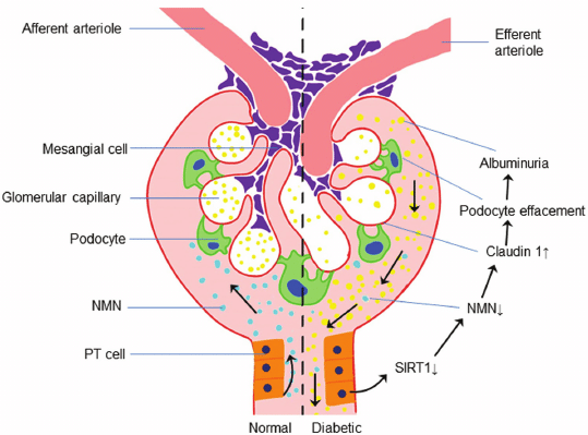
Podocytes are post-mitotic cells with a very long life span, which makes them particularly vulnerable to injury. Podocyte injury underlies many forms of kidney disease, including diabetic nephropathy, focal segmental glomerulosclerosis (FSGS) and human immunodeficiency virus (HIV)-associated nephropathy.
The podocyte cell body has an elaborate cellular architecture. It is divided into four regions known as quarters. A quarter has two rims—an outer rim with foot processes that extend into Bowman’s space and an inner rim that faces the glomerular capillary loop (Figure 1). Each podocyte cell has four quarters—a
What are podocytes cells?
Podocytes are kidney cells that form a key part of the filtration system. They also have a role in preventing the leakage of blood proteins into urine.
These cells take their name from the foot-like processes, called pedicels, which wrap around capillaries in the glomerular filtration barrier, forming slits that allow water and small molecules to pass from the blood into the urine.
Podocytes have been implicated in many kidney diseases, including diabetic nephropathy and focal segmental glomerulosclerosis (FSGS).
Podocytes are cells that make up the glomerulus (the filtering unit of the kidney). They have foot processes that touch the capillary endothelium. They are separated from each other by slit pores.
The podocyte cell is an epithelial cell in the kidneys. It is part of the glomerulus, which filters blood and creates urine. The podocyte has foot processes called pedicels (also known as villi) that wrap around capillaries in the glomerulus. These foot processes create a funnelshaped meshwork that filters blood to produce urine. In addition, they prevent large molecules from passing through the filtration membrane and back into the blood stream.
Podocytes are cells that line the inside of the glomerular capillaries in the kidney. Each podocyte has long processes that wrap around the capillary, forming what is known as a filtration barrier. This barrier allows small molecules and water to leave the blood and enter into Bowman’s space, which leads to a fluid called glomerular filtrate. This fluid is then processed by other parts of the kidney to form urine.
Podocytes are cells that have long processes (foot processes) that wrap around capillaries of the kidney’s glomerulus. The process is called glomerular filtration. The podocytes have holes in them to allow fluids and solutes to pass through into Bowman’s space and then into the tubules of the kidney where they are further processed by the tubule cells.
Podocytes are differentiated epithelial cells that form the filtration barrier in the glomerulus. They have processes that wrap around capillaries to create a barrier for plasma filtration. Podocytes have foot processes that form slit pores through which plasma can pass but where proteins are retained. Podocytes are named for their foot processes, which resemble a bunch of grapes in appearance.
During fetal life, the glomerular filtration barrier is not fully developed. At birth and during the first weeks of life, podocytes continue to mature. The slit diaphragm system becomes more complex with age and a greater number of filtration slits are formed between adjacent podocytes. It is believed that this maturation process may be important in establishing the fine-tuning of filtration throughout the human lifespan.
The high density of slit diaphragms (200–2000 per square micron) in the glomerulus provides a unique pore structure for the control of ultrafiltration. This structure consists of two types of pores:
Fenestrations—Fenestrations range from 50 to 100 nm in diameter. They are large enough to allow free passage of small plasma proteins and permit selective filtration of molecules.[4]
Slit pores—Slit pores are interdigitating extensions from adjacent podocytes that do not reach completely across the specialized basement membrane known as the glomerular basement membrane (GBM).